Category: blogs

  • Breastfeeding: A Natural Defense Against Hormone-Receptor-Negative Breast Cancer

    This being a Breastfeeding week, let’s learn something on the relationship between Cancer and Breastfeeding. Did you know that women who breastfeed have a lower risk of breast cancer especially before menopause, than women who don’t breastfeed?

    Many women are not aware that breastfeeding their babies can help lower their own risk of developing breast cancer; despite the fact that most parents are aware that breastfeeding is the best way to nurture babies and offers them vital health benefits.

    Breastfeeding may be more strongly linked to a lower risk of estrogen receptor-negative (Some breast cancer cells require progesterone and/or oestrogen. Hormone receptors are unique proteins found inside these cancer cells. When hormones attach to hormone receptors, the cancer cells with these receptors grow.

    Hormone receptor-negative tumors are estrogen receptor-negative (ER-negative) and progesterone receptor-negative (PR- negative). Hormone receptors are not expressed in these tumors. Because of this, they either have few or no hormone receptors.

    Approximately 75% to 85% of newly diagnosed breast cancers are hormone receptor-positive, these hormones cane be treated with hormone therapies; hormone theraphy drugs include aromatase inhibitors, tamoxifen, anastrozole, exemestane and letrozole. Hormone receptor-negative breast cancers are not treated with hormone therapies because they don’t have hormone receptors.

    Research suggests that breastfeeding women are less likely to develop pre and post-menopausal breast cancer symptoms. And breastfeeding longer than six months can have additional benefits. Most women ignore their breast health, which results in breast cancer, due to which the number of increased breast cancer cases worldwide.

    Breastfeeding protects children from cancer, it reduces not only the mother’s chances of cancer but also the child’s. It is therefore beneficial to both mother and child. Studies show that breastfeeding prevents the child from being overweight, that later reduces the risk of many cancers such as pancreatic, post-menopausal breast, endometrial, oesophagal, rectal, and kidney cancers. 

    A mother’s hormones are altered by breastfeeding, which may delay the onset of her menstrual cycle and minimize her lifetime exposure to oestrogen and other hormones that may increase her risk of breast cancer. Breastfeeding may also aid in preventing breast cancer-causing mutations in cells.

    Breast cancer can be detected during breastfeeding; when a woman feels a lump in the breast during breastfeeding, it could be a sign of breast cancer, and the lumps can disappear after some time but can translate to cancer as a result of mastitis, which is an infection of the breast tissue by a bacteria which blocks milk ducts. Symptoms are swelling and pain in the breast, skin warmth and redness, fever. Fibro adenomas, which are lumps that feel like marble when touched and always move beneath the skin can also be the symptoms.

    REFERENCES

    Fei, F., Siegal, G. P., & Wei, S. (2022). Characterizing clinicopathologic features of estrogen receptor-positive/progesterone receptor-negative breast cancers. Clinical Breast Cancer, 22(7), e788-e797.

    Huang, Y., Ouyang, Y. Q., & Redding, S. R. (2019). Previous breastfeeding experience and its influence on breastfeeding outcomes in subsequent births: a systematic review. Women and Birth, 32(4), 303-309.

    Kay, J. E., Cardona, B., Rudel, R. A., Vandenberg, L. N., Soto, A. M., Christiansen, S., … & Fenton, S. E. (2022). Chemical effects on breast development, function, and cancer risk: existing knowledge and new opportunities. Current Environmental Health Reports, 9(4), 535-562.

    Rozeboom, B., Dey, N., & De, P. (2019). ER+ metastatic breast cancer: Past, present, and a prescription for an apoptosis-targeted future. American journal of cancer research, 9(12), 2821.

    Skilton, M. R. (2022). Obesity and Cancer. Clinical Obesity in Adults and Children, 211-220.

    Trabert, B., Sherman, M. E., Kannan, N., & Stanczyk, F. Z. (2020). Progesterone and breast cancer. Endocrine reviews, 41(2), 320-344.

  • Improving Breast Cancer Outcomes in Kenya: Focus on Early Detection & Prevention

    This month provides an opportunity for us all to focus on breast cancer and its impact on those affected by the disease in our community.

    The total breast cancer cases notified by county of usual residence between the years 2020 and 2023 amounted to 4,824 people in Kenya, Nairobi County leading with the highest number of diagnosed people at 550 followed by Nakuru County which registered 398 people.

    Finding breast cancer early provides the best chance of surviving the disease. While mammograms can help to detect cancer before you can feel a lump, breast self-exams help to be familiar with how your breasts look and feel so you can alert your doctor if there are any changes.

    Take the time to ‘get to know’ how your breasts normally look and feel through normal regular activities such as showering, getting dressed, using body lotion or looking in the mirror.

    You don’t need to use a special technique, but ensure you look at and feel your breasts regularly. Make sure this includes all parts of your breast, your armpit and up to your collarbone.

    For women of all ages, it is recommended that you be breast aware.

    Changes to look for include:

    • a new lump or lumpiness, especially if it’s only in one breast
    • a change in the size or shape of your breast
    • a change to the nipple, such as crusting, ulcer, redness or inversion
    • a nipple discharge that occurs without squeezing
    • a change in the skin of your breast such as redness or dimpling
    • An unusual pain that doesn’t go away.

    Visit a doctor in case you notice any changes in your breast.

    Breast cancer signs

    According to doctors, it is quite difficult for breast cancer to get diagnosed for women under 40 because their breasts are denser. This means that a tumour, if present, is not likely to show up in a mammogram.

    The most common way to detect breast cancer in younger women is to keep a close check on the changes in your breasts. In fact, according to Healthline, a majority of young women diagnosed with breast cancer discover an abnormality themselves. If you notice any changes in your breast, like changes in the skin, nipple discharge, pain, tenderness, or a lump or mass in the breast or underarm area, please report it immediately to a doctor.

    Breast cancer is one of the most common cancers in women and there is a misconception among people that it only happens to older women. While it’s true that most breast cancers are found in women who are 40 years or older, it is imperative to know that it can strike any female at any age.

    Pain in any Area of The Breast

    Check for pain and tenderness in any area of your breast. While lumps don’t usually hurt, some may cause a prickly sensation. However, pain in your breasts can also be due to numerous other benign reasons and hormonal changes. 

    Unusual Nipple Discharge

    An unusual nipple discharge that’s clear, red, brown, or yellow (other than breast milk) can be an early sign of breast cancer. If you notice this, consult a doctor for an evaluation.

    Breast Changes

    Look for changes in shape, swelling, and difference in size, texture, or temperature of either one or both breasts.

    Unexplained Redness/Rash

    Early warning signs of breast cancer can include unexplained redness, swelling, itchiness, or rash on one or both breasts.

    Swelling around the Collarbone or Armpit

    Consult your doctor if you notice swelling around your collarbone or under the arm. It could be a sign that the breast cancer has spread to the lymph nodes in that area.

    While these are some of the early signs of breast cancer, it is important to remember that these can also be caused by benign conditions. It’s always best to consult a doctor so that they can evaluate and help you determine whether it is cause for concern.

    Apart from this, it’s also important to keep in mind that often, women with breast cancer have no symptoms. This is why regular breast screening is important to detect it. The main tool to diagnose breast cancer is a mammogram, which basically shows normal and abnormal breast tissue.

    It is recommended that women should start getting mammograms when they cross the age of 40 and should continue getting one every 1 or 2 years. Meanwhile, for women above the age of 20, monthly breast self-examination is recommended.

  • Breast Cancer Status in Kenya – National Cancer Registry of Kenya (NaCaRe-KE)

    The National Cancer Registry of Kenya (NaCaRe-KE) was established in 2021 and launched in the same year.  The first report on the status of cancer in Kenya, utilized 6,778 records for the period July 2021 to June 2022, notified from 42 facilities across 21 counties.  From the report, it is established that three most common cancers are breast (15.1%), cervix (13.3%) and oesophagus (11.8%). 

    The total breast cancer cases notified by county of usual residence between the years 2020 and 2023 amounted to 4,824 people, Nairobi County leading with the highest number of diagnosed people at 550 followed by Nakuru County which registered 398 people. Counties that diagnosed the least number of breast cancer as notified to NaCaRe-KE included Turkana, Tana River and Wajir that registered 3, 2 and 1 cases respectively as shown in table 1 below.

    Table 1: Distribution by County of Usual Residence per Year – Filtered by Date of diagnosis

    County of Usual Residence2020202120222023TOTAL
     Nairobi County            1          51         354         144         550
     Nakuru County          96        105         130           67         398
     Nyeri County           96         179           94         369
     Kiambu County           14         199           70         283
     Machakos County           30         112           77         219
     Mombasa County            4          20           23         143         190
     Meru County          61          27           65           24         177
     Makueni County             8           96           49         153
     Kirinyaga County            1          33           69           49         152
     Muranga County             6         111           35         152
     Embu County            2          36           75           27         140
     Kitui County          22          19           59           32         132
     Kakamega County           23           65           40         128
     Kajiado County             9           47           36           92
     Laikipia County          10          13           36           33           92
     Nyandarua County          10          17           39           15           81
     Kisumu County           15           37           28           80
     Kilifi County            4            9            7           59           79
     Kisii County            1            7           37           30           75
     Siaya County           10           26           22           58
     Uasin Gishu County            1          29           21            7           58
     Taita Taveta County           13           21           19           53
     Bungoma County           16           16           14           46
     Busia County           15           17           11           43
     Vihiga County             8           15           20           43
     Nandi County             5           13           24           42
     Kwale County            1            3            4           26           34
     Baringo County            6          12            4            5           27
     Homa Bay County            1            5            9           12           27
     Migori County             3           15            9           27
     Tharaka Nithi County             6           15            5           26
     Trans Nzoia County             9            8            9           26
     Kericho County            1            8           10            5           24
     Nyamira County             4            6           14           24
     Bomet County           10           10            2           22
     Narok County             6            5            9           20
     Garissa County             4            8            1           13
     Lamu County            1            2            1            6           10
     Isiolo County              5            2            7
     Marsabit County             2            1            3            6
     West Pokot County             2            2            2            6
     Elgeyo Marakwet County             2            2            1            5
     Mandera County              5             5
     Samburu County            1             2            1            4
     Turkana County             1            1            1            3
     Tana River County              1            1            2
     Wajir County              1             1
     Non-resident             16            4           20
     Missing        357        207           33            3         600
     TOTAL        581        920     2,033     1,290     4,824

    Distribution by County of Notification per Year – Filtered by Date of Diagnosis

    Distribution by County of notification means the county in which the cancer case was diagnosed. From table 2 below, it is noted that Nairobi County leads by 2150 of cases notified; meaning therefore that most people seek cancer medication at the capital city.

    Table 2: Distribution by County of Notification per Year – Filtered by Date of Diagnosis

    County of Usual Res 2020 2021 2022 2023 TOTAL
     Nairobi County        218        215      1,288         429      2,150
     Nakuru County        128        126         124           82         460
     Nyeri County         117         143         125         385
     Machakos County        101          73           78           65         317
     Mombasa County          18          31            7         242         298
     Uasin Gishu County         116           79           36         231
     Kakamega County           89           49           42         180
     Kisumu County           34           42           71         147
     Meru County          61          32           25           23         141
     Embu County            3          47           65           24         139
     Makueni County             56           19           75
     Kitui County          22          13           17           14           66
     Bomet County          20          11           13            4           48
     Taita Taveta County           13           15           13           41
     Kiambu County              4           31           35
     Kisii County              2           28           30
     Nandi County              3           24           27
     Garissa County          10            3            8            2           23
     Laikipia County              5            5           10
     Muranga County             10            10
     Kirinyaga County               8            8
     Vihiga County               3            3
     TOTAL        581        920      2,033      1,290      4,824

    In terms of distribution of cancer type in the breast (primary site), Breast NOS (excludes Skin of breast C44.5) accounts for 94.44% of the all cancers of the breast; 4556 of the total 4824 as notified between 2020 and 2023. Breast NOS in simple terms is the Invasive Ductal Carcinoma of Breast, Not Otherwise Specified (NOS), they are invasive breast cancers, which have a tendency to be aggressive and spread. This subset comprises the largest group and also the most common type of breast cancers as seen in table 2 below.

    Table 3: Distribution of Cancer Type by /year – Filtered by Date of Diagnosis:

    Primary Site 2020 2021 2022 2023 TOTAL
     Breast NOS (excludes Skin of breast C44.5)        542        893     1,957     1,164     4,556
     Upper outer quadrant of breast          10          12           20           27           69
     Central portion of breast            2            1           20           28           51
     Upper inner quadrant of breast            3            5            7           19           34
     Overlapping lesion of breast          19            6            6            3           34
     Axillary tail of breast             11           17           28
     Lower inner quadrant of breast            4             9            8           21
     Nipple             3            1           15           19
     Lower outer quadrant of breast            1             2            9           12
     TOTAL        581        920     2,033     1,290     4,824

    In Kenya, the majority of breast cancer cases discovered are at stage IV, accounting for 37.2% of all cases as seen in table 4 below. This is the leading factor towards mortalities as late discoveries reduces the likelihood of survival. Early screening of breast cancer increases the chances of managing the disease through prompt treatment and care, therefore increasing the chances of survival.

    Table 4: Distribution by Stage at Diagnosis per Year – Filtered by Date of Diagnosis:

    Stage at Diagnosis2020202120222023TOTALPercentage
    In situ 22036582.6%
    Stage I121856291155.1%
    Stage II256723521053723.8%
    Stage III4414425626470831.4%
    Stage IV5617137224184037.2%
    TOTAL1374029397802258100.0%
  • Understanding Brain Tumors: A Guide for Patients

    A brain tumor is a growth of cells in the brain or near it. Brain tumors can happen in the brain tissue. Brain tumors also can happen near the brain tissue. Nearby locations include nerves, the pituitary gland, the pineal gland, and the membranes that cover the surface of the brain.

    Brain tumors can begin in the brain. These are called primary brain tumors. Sometimes, cancer spreads to the brain from other parts of the body. These tumors are secondary brain tumors, also called metastatic brain tumors.

    Many different types of primary brain tumors exist. Some brain tumors aren’t cancerous. These are called noncancerous brain tumors or benign brain tumors. Noncancerous brain tumors may grow over time and press on the brain tissue. Other brain tumors are brain cancers, also called malignant brain tumors. Brain cancers may grow quickly. The cancer cells can invade and destroy the brain tissue.

    Brain tumors range in size from very small to very large. Some brain tumors are found when they are very small because they cause symptoms that you notice right away. Other brain tumors grow very large before they’re found. Some parts of the brain are less active than others. If a brain tumor starts in a part of the brain that’s less active, it might not cause symptoms right away. The brain tumor size could become quite large before the tumor is detected.

    Brain tumor treatment options depend on the type of brain tumor you have, as well as its size and location. Common treatments include surgery and radiation therapy.

    Types

    There are many types of brain tumors. The type of brain tumor is based on the kind of cells that make up the tumor. Special lab tests on the tumor cells can give information about the cells. Your health care team uses this information to figure out the type of brain tumor.

    Some types of brain tumors usually aren’t cancerous. These are called noncancerous brain tumors or benign brain tumors. Some types of brain tumors usually are cancerous. These types are called brain cancers or malignant brain tumors. Some brain tumor types can be benign or malignant.

    Benign brain tumors tend to be slow-growing brain tumors. Malignant brain tumors tend to be fast-growing brain tumors.

    Types of brain tumors include:

    • Gliomas and related brain tumors. Gliomas are growths of cells that look like glial cells. The glial cells surround and support nerve cells in the brain tissue. Types of gliomas and related brain tumors include astrocytoma, glioblastoma, oligodendroglioma and ependymoma. Gliomas can be benign, but most are malignant. Glioblastoma is the most common type of malignant brain tumor.
    • Choroid plexus tumors. Choroid plexus tumors start in cells that make the fluid that surrounds the brain and spinal cord. This fluid is called cerebrospinal fluid. Choroid plexus tumors are located in the fluid-filled cavities in the brain, called the ventricles. Choroid plexus tumors can be benign or malignant. Choroid plexus carcinoma is the malignant form of this type of brain tumor. It’s more common in children.
    • Embryonal tumors. Embryonal tumors begin in cells that are left over from fetal development. The cells, called embryonal cells, stay in the brain after birth. Embryonal tumors are malignant brain tumors that happen most often in babies and young children. The most common type of embryonal tumor is medulloblastoma. It’s usually located in the lower back part of the brain, called the cerebellum.
    • Germ cell tumors. Germ cell tumors start in reproductive cells, called germ cells, that go on to become the sperm and egg cells. Germ cells are mostly in the ovaries and testicles. But sometimes they’re in other parts of the body, including the brain. When germ cell tumors happen in the brain, they’re often located near the pineal gland or the pituitary gland. Germ cell tumors are mostly benign. They’re more common in children.
    • Pineal tumors. Pineal tumors start in and around the brain’s pineal gland. The pineal gland is located in the center of the brain. It makes a hormone called melatonin that helps with sleep. Pineal tumors can be benign or malignant. Pineoblastoma is a malignant type of pineal tumor that’s most common in children.
    • Meningiomas. Meningiomas are brain tumors that start in the membranes around the brain and spinal cord. Meningiomas are usually benign, but sometimes they can be malignant. Meningiomas are the most common type of benign brain tumor.
    • Nerve tumors. Nerve tumors are growths that happen in and around nerves. The most common type that happens in the head is acoustic neuroma, also called schwannoma. This benign tumor is located on the main nerve that connects the inner ear to the brain.
    • Pituitary tumors. Brain tumors can begin in and around the pituitary gland. This small gland is located near the base of the brain. Most tumors that happen in and around the pituitary gland are benign. Pituitary tumors happen in the pituitary gland itself. Craniopharyngioma is a type of brain tumor that happens near the pituitary gland.
    • Other brain tumors. Many other types of rare tumors can happen in and around the brain. Tumors can start in the muscles, blood vessels and connective tissue around the brain. Tumors can form in the bones of the skull. Malignant brain tumors can start from the germ-fighting immune system cells in the brain. This type of brain cancer is called primary central nervous system lymphoma.

  • Improving Childhood Cancer Care: Prevention, Diagnosis, and Treatment

    Childhood cancer is a diverse and heterogeneous group of rare cancers that are unified by the age of the cohort, childhood cancer is defined as the group of cancer that arises between birth and 19 years of age.

    Care for cancer, like so many other diseases, reflects the inequalities and inequities in our societies. The impact on children is devastating. Each year, an estimated 400 000 children are diagnosed with cancer around the world. The vast majority of these children live in low- and middle-income countries, where treatment is often unavailable, unaffordable or of poor quality. The survival rate for these children is estimated to be between 15% and 45%. This compares to a survival rate of more than 80% in high-income countries.

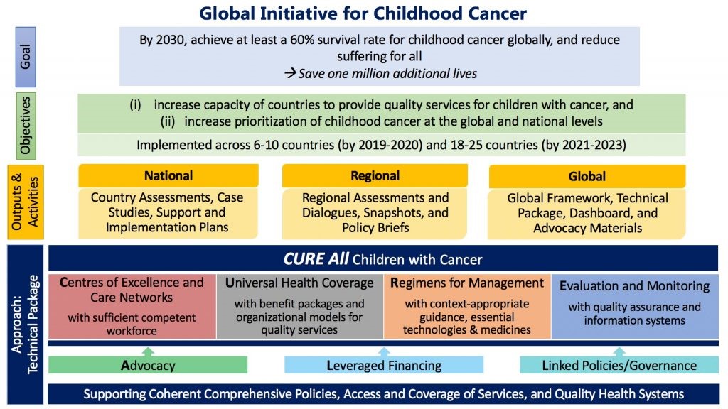
    We continue to raise awareness about childhood cancer and express support for children and adolescents with cancer, survivors and their families. This is in line with WHO Global Initiative on Childhood Cancer, which aims at reaching at least a 60% survival rate for children with cancer by 2030.

    This new target represents a doubling of the global cure rate for children with cancer. The aims of the Initiative are two-fold: to increase prioritization of childhood cancer through awareness raising at global and national levels and to expand the capacity of countries to deliver best practice in childhood cancer care. 

    The institute is working to improve primary prevention of childhood cancers through:

    • Mitigation of risk factors and their determinants
    • Enhancing access to appropriate cancer screening services for early detection
    • Strengthening the referral pathway for patients with cancer and those suspected to have cancer
    • Promotion of access to optimal diagnostics, treatment, palliative care and survivorship
    • Strengthening cancer information systems, registration and surveillance

    This can be achieved through multi-sectorial approach which will involve both health and non-health sectors. It will also involve the recognition of the best practices and scientific evidence supported through monitoring and evaluation and research.

    The types of cancers that occur most often in children are different from those seen in adults. The most common cancers of children are:

    • Leukemia
    • Brain and spinal cord tumors
    • Neuroblastoma
    • Wilms tumor
    • Lymphoma (including both Hodgkin and non-Hodgkin)
    • Rhabdomyosarcoma
    • Retinoblastoma
    • Bone cancer (including osteosarcoma and Ewing sarcoma)

  • Gastrointestinal Stromal Tumors (GISTs): Types, Causes, and Risk Factors

    A gastrointestinal stromal tumor (GIST) is a type of tumor that occurs in the gastrointestinal tract, most commonly in the stomach or small intestine. This type of tumor is thought to grow from specialized cells found in the gastrointestinal tract called interstitial cells of Cajal (ICCs) or precursors to these cells. Affected individuals can develop one or more tumors. GISTs are usually found in adults between ages 40 and 70; rarely, children and young adults develop this type of tumor.

    Small tumors may cause no signs or symptoms. However, some people with GISTs may experience pain or swelling in the belly area (abdomen), nausea, vomiting, loss of appetite, or weight loss. Sometimes, tumors cause bleeding into the gastrointestinal tract, which may lead to low red blood cell counts and, consequently, weakness and tiredness. Bleeding into the intestines may cause black and tarry stools, and bleeding into the throat or stomach may cause vomiting of blood.

    Affected individuals with no family history of GIST typically have only one tumor (called a sporadic GIST). People with a family history of GISTs (called familial GISTs) often have multiple tumors and additional signs or symptoms, including noncancerous overgrowth (hyperplasia) of other cells in the gastrointestinal tract and patches of dark skin on various areas of the body. Some affected individuals have a skin condition called urticaria pigmentosa (also known as maculopapular cutaneous mastocytosis), which is characterized by raised patches of brownish skin that sting or itch when touched.

    A rare form of GIST, called succinate dehydrogenase (SDH)-deficient GIST, tends to occur in childhood or young adulthood and affects females more commonly than males. In this form, tumors are almost always in the stomach. Individuals with an SDH-deficient GIST have a high risk of developing other types of tumors, particularly noncancerous tumors in the nervous system called paragangliomas and noncancerous lung tumors called pulmonary chondromas. When GISTs occur in combination with paragangliomas, the condition is known as Carney-Stratakis syndrome; the combination of GISTs, paragangliomas, and pulmonary chondromas is known as Carney triad; and the combination of GISTs and pulmonary chondroma is known as incomplete Carney triad.

  • Genetic Risk Factors for Leukemia in Children: Down Syndrome and Other Conditions

    In children and teenagers, leukaemia accounts for roughly one out of every 3 cancer cases. Acute lymphocytic leukaemia (ALL) is the most common kind of leukaemia in kids. Acute myeloid leukaemia (AML) makes up the majority of the remaining cases. In children, chronic leukaemias are uncommon.

    A few factors are known to increase the risk of childhood leukaemia.

    Genetic Risk Factors

    Our DNA, which makes up our genes, contains risk factors that are genetic in nature. They are frequently passed on from our parents. While some genetic predispositions raise the incidence of paediatric leukaemia, the majority of leukaemias have no known genetic basis.

    Genetic Syndromes

    Some genetic disorders increase a child’s risk of developing leukemia:

    Down syndrome (trisomy 21): Children with Down syndrome have an extra copy of chromosome 21. They are many times more likely to develop either acute lymphocytic leukemia (ALL) or acute myeloid leukemia (AML) than are other children, with an overall risk of about 2% to 3%. Down syndrome has also been linked with transient leukemia (also known as transient myeloproliferative disorder) – a leukemia-like condition within the first month of life, which often resolves on its own without treatment.

    Li-Fraumeni syndrome: This is a rare inherited condition caused by a change in the TP53 gene. People with this change have a higher risk of developing several kinds of cancer, including leukemia, bone or soft tissue sarcomas, breast cancer, adrenal gland cancer, and brain tumors.

    Leukaemia risk is also elevated in other genetic illnesses (including neurofibromatosis and Fanconi anaemia), as well as in several other malignancies.

    Inherited Immune System Problems

    Certain inherited conditions cause children to be born with immune system problems. These include:

    • Ataxia-telangiectasia
    • Wiskott-Aldrich syndrome
    • Bloom syndrome
    • Shwachman-Diamond syndrome

    Along with an increased risk of getting serious infections from reduced immune defenses, these children might also have an increased risk of leukemia.

    Lifestyle-Related Risk Factors

    Smoking, being overweight, consuming excessive amounts of alcohol, and being exposed to the sun too much are all lifestyle-related risk factors for several adult malignancies. However, they are unlikely to have an impact on the majority of children malignancies. These types of characteristics are significant in many adult cancers.

    Although not all studies have established a correlation, some have suggested that a pregnant woman who drinks a lot of alcohol may put her unborn child at risk for leukaemia.

    Environmental Risk Factors

    Environmental risk factors are influences in our surroundings, such as radiation and certain chemicals that increase the risk of getting diseases such as leukemias.

    Radiation Exposure

    Exposure to high levels of radiation is a risk factor for childhood leukemia. Japanese atomic bomb survivors had a greatly increased risk of developing AML. If a fetus is exposed to radiation within the first months of development, there may also be an increased risk of childhood leukemia, but the extent of the risk is not clear.

    The possible risks from fetal or childhood exposure to lower levels of radiation, such as from x-ray tests or CT scans, are not known for sure. Some studies have found a slight increase in risk, while others have found no increased risk. Any risk increase is likely to be small, but to be safe, most doctors recommend that pregnant women and children not get these tests unless they are absolutely needed.

    Exposure to Chemotherapy and Certain other Chemicals

    Children and adults treated for other cancers with certain chemotherapy drugs have a higher risk of getting a second cancers, usually AML, later in life. Drugs such as cyclophosphamide, doxorubicin, etoposide, and teniposide have been linked to a higher risk of leukemia. These leukemias usually develop within 5 to 10 years of treatment, and they tend to be hard to treat.

    Exposure to chemicals such as benzene (a solvent used in the cleaning industry and to manufacture some drugs, plastics, and dyes) may cause acute leukemia in adults and, rarely, in children. Chemical exposure is more strongly linked to an increased risk of AML than to ALL.

    A number of studies have discovered a potential connection between pesticide exposure in the home during pregnancy or the early years of life and childhood leukaemia. There may be a higher probability among moms who are exposed to pesticides at work before giving birth, according to some research.

    Immune System Suppression

    Children who are getting intensive treatment to suppress their immune system (mainly children who have had organ transplants) have an increased risk of certain cancers, such as lymphoma and ALL.

  • Understanding Brain Tumors: A Comprehensive Guide

    A brain tumor is a growth of cells in the brain or near it. Brain tumors can happen in the brain tissue. Brain tumors also can happen near the brain tissue. Nearby locations include nerves, the pituitary gland, the pineal gland, and the membranes that cover the surface of the brain.

    Brain tumors can begin in the brain. These are called primary brain tumors. Sometimes, cancer spreads to the brain from other parts of the body. These tumors are secondary brain tumors, also called metastatic brain tumors.

    Many different types of primary brain tumors exist. Some brain tumors aren’t cancerous. These are called noncancerous brain tumors or benign brain tumors. Noncancerous brain tumors may grow over time and press on the brain tissue. Other brain tumors are brain cancers, also called malignant brain tumors. Brain cancers may grow quickly. The cancer cells can invade and destroy the brain tissue.

    Brain tumors range in size from very small to very large. Some brain tumors are found when they are very small because they cause symptoms that you notice right away. Other brain tumors grow very large before they’re found. Some parts of the brain are less active than others. If a brain tumor starts in a part of the brain that’s less active, it might not cause symptoms right away. The brain tumor size could become quite large before the tumor is detected.

    Brain tumor treatment options depend on the type of brain tumor you have, as well as its size and location. Common treatments include surgery and radiation therapy.

    Types

    There are many types of brain tumors. The type of brain tumor is based on the kind of cells that make up the tumor. Special lab tests on the tumor cells can give information about the cells. Your health care team uses this information to figure out the type of brain tumor.

    Some types of brain tumors usually aren’t cancerous. These are called noncancerous brain tumors or benign brain tumors. Some types of brain tumors usually are cancerous. These types are called brain cancers or malignant brain tumors. Some brain tumor types can be benign or malignant.

    Benign brain tumors tend to be slow-growing brain tumors. Malignant brain tumors tend to be fast-growing brain tumors.

    Types of brain tumors include:

    • Gliomas and related brain tumors. Gliomas are growths of cells that look like glial cells. The glial cells surround and support nerve cells in the brain tissue. Types of gliomas and related brain tumors include astrocytoma, glioblastoma, oligodendroglioma and ependymoma. Gliomas can be benign, but most are malignant. Glioblastoma is the most common type of malignant brain tumor.
    • Choroid plexus tumors. Choroid plexus tumors start in cells that make the fluid that surrounds the brain and spinal cord. This fluid is called cerebrospinal fluid. Choroid plexus tumors are located in the fluid-filled cavities in the brain, called the ventricles. Choroid plexus tumors can be benign or malignant. Choroid plexus carcinoma is the malignant form of this type of brain tumor. It’s more common in children.
    • Embryonal tumors. Embryonal tumors begin in cells that are left over from fetal development. The cells, called embryonal cells, stay in the brain after birth. Embryonal tumors are malignant brain tumors that happen most often in babies and young children. The most common type of embryonal tumor is medulloblastoma. It’s usually located in the lower back part of the brain, called the cerebellum.
    • Germ cell tumors. Germ cell tumors start in reproductive cells, called germ cells, that go on to become the sperm and egg cells. Germ cells are mostly in the ovaries and testicles. But sometimes they’re in other parts of the body, including the brain. When germ cell tumors happen in the brain, they’re often located near the pineal gland or the pituitary gland. Germ cell tumors are mostly benign. They’re more common in children.
    • Pineal tumors. Pineal tumors start in and around the brain’s pineal gland. The pineal gland is located in the center of the brain. It makes a hormone called melatonin that helps with sleep. Pineal tumors can be benign or malignant. Pineoblastoma is a malignant type of pineal tumor that’s most common in children.
    • Meningiomas. Meningiomas are brain tumors that start in the membranes around the brain and spinal cord. Meningiomas are usually benign, but sometimes they can be malignant. Meningiomas are the most common type of benign brain tumor.
    • Nerve tumors. Nerve tumors are growths that happen in and around nerves. The most common type that happens in the head is acoustic neuroma, also called schwannoma. This benign tumor is located on the main nerve that connects the inner ear to the brain.
    • Pituitary tumors. Brain tumors can begin in and around the pituitary gland. This small gland is located near the base of the brain. Most tumors that happen in and around the pituitary gland are benign. Pituitary tumors happen in the pituitary gland itself. Craniopharyngioma is a type of brain tumor that happens near the pituitary gland.
    • Other brain tumors. Many other types of rare tumors can happen in and around the brain. Tumors can start in the muscles, blood vessels and connective tissue around the brain. Tumors can form in the bones of the skull. Malignant brain tumors can start from the germ-fighting immune system cells in the brain. This type of brain cancer is called primary central nervous system lymphoma.
  • Colorectal Risk Factors you can Change

    Many lifestyle-related factors have been linked to colorectal cancer. In fact, more than half of all colorectal cancers are linked to risk factors that can be changed. These include:

    • Being overweight or obese
    • Diabetes mellitus, Type 2
    • Certain types of diets – A long-term diet that’s high in red meats (such as beef, pork, lamb, or liver) and processed meats (like hot dogs and some lunch meats) raises your colorectal cancer risk.
    • Having a low blood level of vitamin D may also increase your risk.
    • Smoking
    • Alcohol use

    If you are overweight or obese (very overweight), your risk of developing and dying from colorectal cancer is higher. Being overweight raises the risk of colorectal cancer in people, but the link seems to be stronger in men. Getting to and staying at a healthy weight may help lower your risk.

    People with type 2 diabetes mellitus are more likely than people who don’t to develop colorectal cancer. Researchers suspect that this higher risk may be due to high levels of insulin in people with diabetes mellitus. Both type 2 diabetes and colorectal cancer share some of the same risk factors (such as being overweight and physical inactivity). But even after taking these factors into account, people with type 2 diabetes still have an increased risk. They also tend to have a less favorable prognosis (outlook) after diagnosis.

    Cooking meats at very high temperatures (frying, broiling, or grilling) creates chemicals that might raise your cancer risk.

    Following a healthy eating pattern that includes plenty of fruits, vegetables, and whole grains, and that limits or avoids red and processed meats and sugary drinks probably lowers risk.

    People who have smoked tobacco for a long time are more likely to develop and die from colorectal cancer than people who don’t smoke. Smoking tobacco also increases the risk for people to develop colon polyps. Smoking is a well-known cause of lung cancer, but it’s linked to a lot of other cancers, too.

  • Breast Cancer Status in Kenya – National Cancer Registry of Kenya (NaCaRe-KE)

    The National Cancer Registry of Kenya (NaCaRe-KE) was established in 2021 and launched in the same year.  The first report on the status of cancer in Kenya, utilized 6,778 records for the period July 2021 to June 2022, notified from 42 facilities across 21 counties.  From the report, it is established that three most common cancers are breast (15.1%), cervix (13.3%) and oesophagus (11.8%). 

    The total breast cancer cases notified by county of usual residence between the years 2020 and 2023 amounted to 4,824 people, Nairobi County leading with the highest number of diagnosed people at 550 followed by Nakuru County which registered 398 people. Counties that diagnosed the least number of breast cancer as notified to NaCaRe-KE included Turkana, Tana River and Wajir that registered 3, 2 and 1 cases respectively as shown in table 1 below.

    Table 1: Distribution by County of Usual Residence per Year – Filtered by Date of diagnosis

    County of Usual Residence2020202120222023TOTAL
     Nairobi County            1          51         354         144         550
     Nakuru County          96        105         130           67         398
     Nyeri County           96         179           94         369
     Kiambu County           14         199           70         283
     Machakos County           30         112           77         219
     Mombasa County            4          20           23         143         190
     Meru County          61          27           65           24         177
     Makueni County             8           96           49         153
     Kirinyaga County            1          33           69           49         152
     Muranga County             6         111           35         152
     Embu County            2          36           75           27         140
     Kitui County          22          19           59           32         132
     Kakamega County           23           65           40         128
     Kajiado County             9           47           36           92
     Laikipia County          10          13           36           33           92
     Nyandarua County          10          17           39           15           81
     Kisumu County           15           37           28           80
     Kilifi County            4            9            7           59           79
     Kisii County            1            7           37           30           75
     Siaya County           10           26           22           58
     Uasin Gishu County            1          29           21            7           58
     Taita Taveta County           13           21           19           53
     Bungoma County           16           16           14           46
     Busia County           15           17           11           43
     Vihiga County             8           15           20           43
     Nandi County             5           13           24           42
     Kwale County            1            3            4           26           34
     Baringo County            6          12            4            5           27
     Homa Bay County            1            5            9           12           27
     Migori County             3           15            9           27
     Tharaka Nithi County             6           15            5           26
     Trans Nzoia County             9            8            9           26
     Kericho County            1            8           10            5           24
     Nyamira County             4            6           14           24
     Bomet County           10           10            2           22
     Narok County             6            5            9           20
     Garissa County             4            8            1           13
     Lamu County            1            2            1            6           10
     Isiolo County              5            2            7
     Marsabit County             2            1            3            6
     West Pokot County             2            2            2            6
     Elgeyo Marakwet County             2            2            1            5
     Mandera County              5             5
     Samburu County            1             2            1            4
     Turkana County             1            1            1            3
     Tana River County              1            1            2
     Wajir County              1             1
     Non-resident             16            4           20
     Missing        357        207           33            3         600
     TOTAL        581        920     2,033     1,290     4,824

    Distribution by County of Notification per Year – Filtered by Date of Diagnosis

    Distribution by County of notification means the county in which the cancer case was diagnosed. From table 2 below, it is noted that Nairobi County leads by 2150 of cases notified; meaning therefore that most people seek cancer medication at the capital city.

    Table 2: Distribution by County of Notification per Year – Filtered by Date of Diagnosis

    County of Usual Res 2020 2021 2022 2023 TOTAL
     Nairobi County        218        215      1,288         429      2,150
     Nakuru County        128        126         124           82         460
     Nyeri County         117         143         125         385
     Machakos County        101          73           78           65         317
     Mombasa County          18          31            7         242         298
     Uasin Gishu County         116           79           36         231
     Kakamega County           89           49           42         180
     Kisumu County           34           42           71         147
     Meru County          61          32           25           23         141
     Embu County            3          47           65           24         139
     Makueni County             56           19           75
     Kitui County          22          13           17           14           66
     Bomet County          20          11           13            4           48
     Taita Taveta County           13           15           13           41
     Kiambu County              4           31           35
     Kisii County              2           28           30
     Nandi County              3           24           27
     Garissa County          10            3            8            2           23
     Laikipia County              5            5           10
     Muranga County             10            10
     Kirinyaga County               8            8
     Vihiga County               3            3
     TOTAL        581        920      2,033      1,290      4,824

    In terms of distribution of cancer type in the breast (primary site), Breast NOS (excludes Skin of breast C44.5) accounts for 94.44% of the all cancers of the breast; 4556 of the total 4824 as notified between 2020 and 2023. Breast NOS in simple terms is the Invasive Ductal Carcinoma of Breast, Not Otherwise Specified (NOS), they are invasive breast cancers, which have a tendency to be aggressive and spread. This subset comprises the largest group and also the most common type of breast cancers as seen in table 2 below.

    Table 3: Distribution of Cancer Type by /year – Filtered by Date of Diagnosis:

    Primary Site 2020 2021 2022 2023 TOTAL
     Breast NOS (excludes Skin of breast C44.5)        542        893     1,957     1,164     4,556
     Upper outer quadrant of breast          10          12           20           27           69
     Central portion of breast            2            1           20           28           51
     Upper inner quadrant of breast            3            5            7           19           34
     Overlapping lesion of breast          19            6            6            3           34
     Axillary tail of breast             11           17           28
     Lower inner quadrant of breast            4             9            8           21
     Nipple             3            1           15           19
     Lower outer quadrant of breast            1             2            9           12
     TOTAL        581        920     2,033     1,290     4,824

    In Kenya, the majority of breast cancer cases discovered are at stage IV, accounting for 37.2% of all cases as seen in table 4 below. This is the leading factor towards mortalities as late discoveries reduces the likelihood of survival. Early screening of breast cancer increases the chances of managing the disease through prompt treatment and care, therefore increasing the chances of survival.

    Table 4: Distribution by Stage at Diagnosis per Year – Filtered by Date of Diagnosis:

    Stage at Diagnosis2020202120222023TOTALPercentage
    In situ 22036582.6%
    Stage I121856291155.1%
    Stage II256723521053723.8%
    Stage III4414425626470831.4%
    Stage IV5617137224184037.2%
    TOTAL1374029397802258100.0%