21st February, 2025
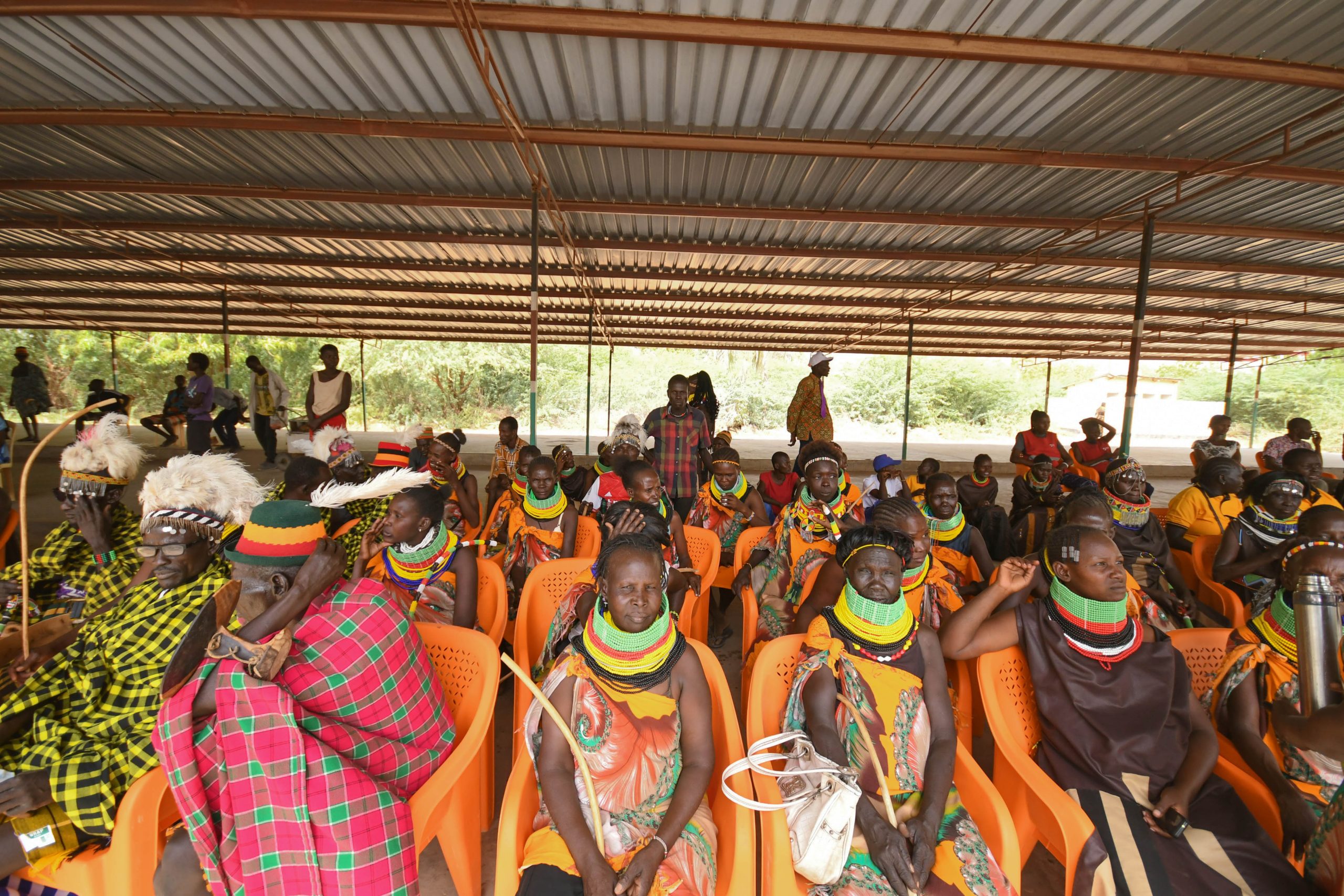
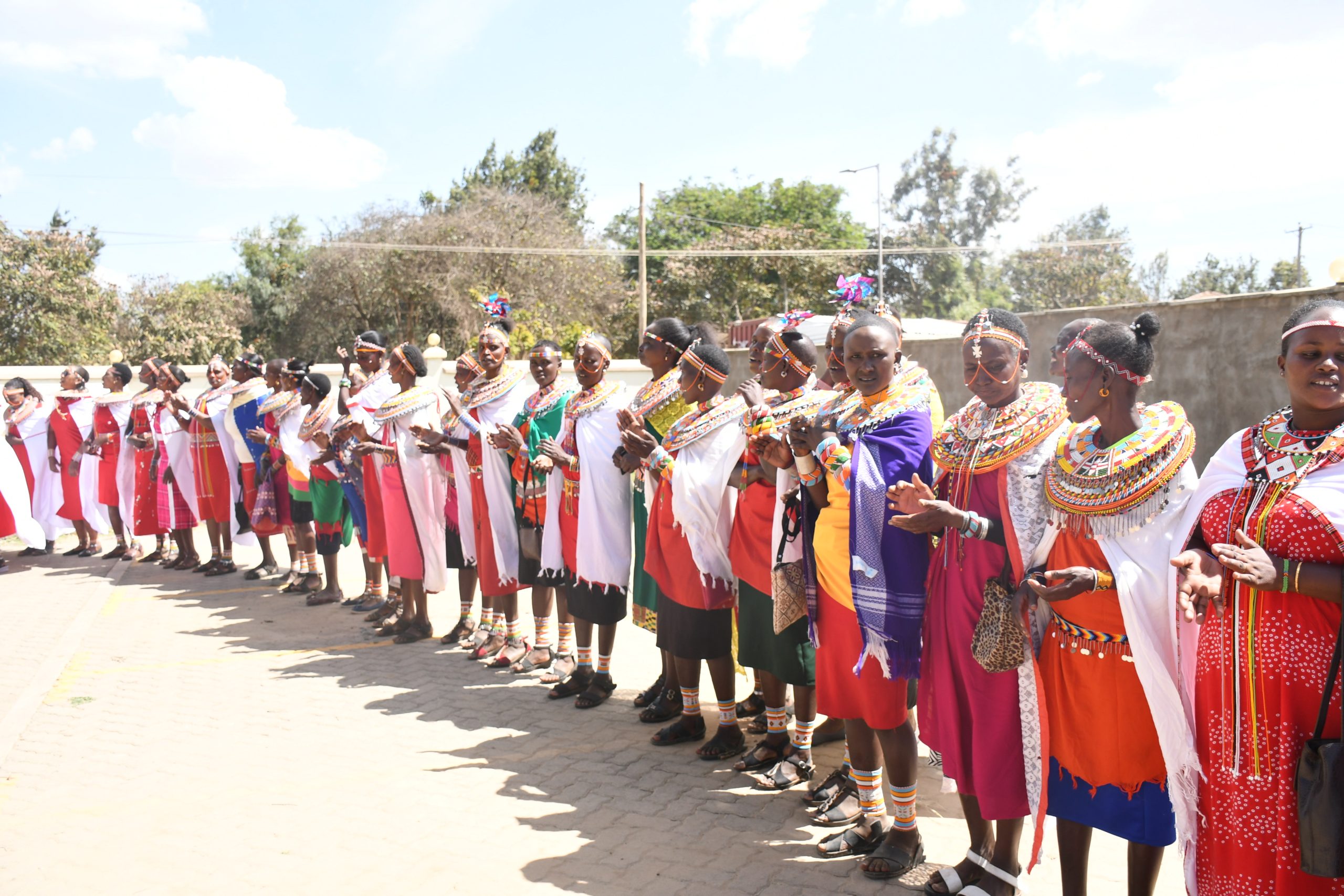
Maralal, Samburu County – Today marked a momentous occasion for the people of Samburu County as the Cabinet Secretary for Health, Dr. Debora Barasa officially launched a state-of-the-art oncology clinic at the Samburu County Referral Hospital. This significant milestone promises to transform cancer care access for residents of this remote region.
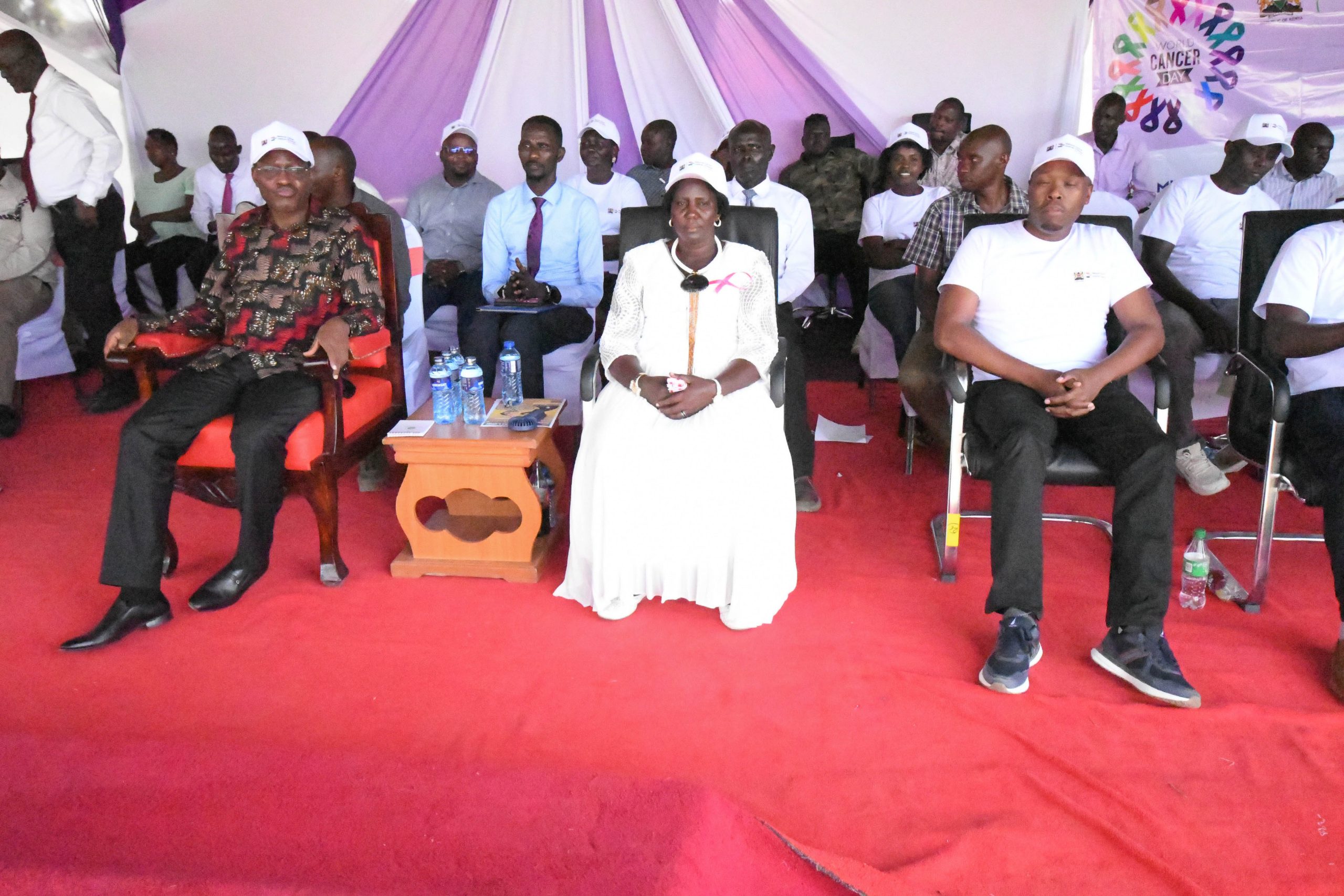
The launch event was a testament to the collaborative spirit driving healthcare advancement in Kenya. The Cabinet Secretary, Dr. Debora Barasa was warmly welcomed by County leadership, NCI-K leadership, including Dr. Timothy Olweny, Board Chair of the National Cancer Institute of Kenya, and Dr. Elias Melly, CEO of NCI-K, and both instrumental in bringing this project to fruition.
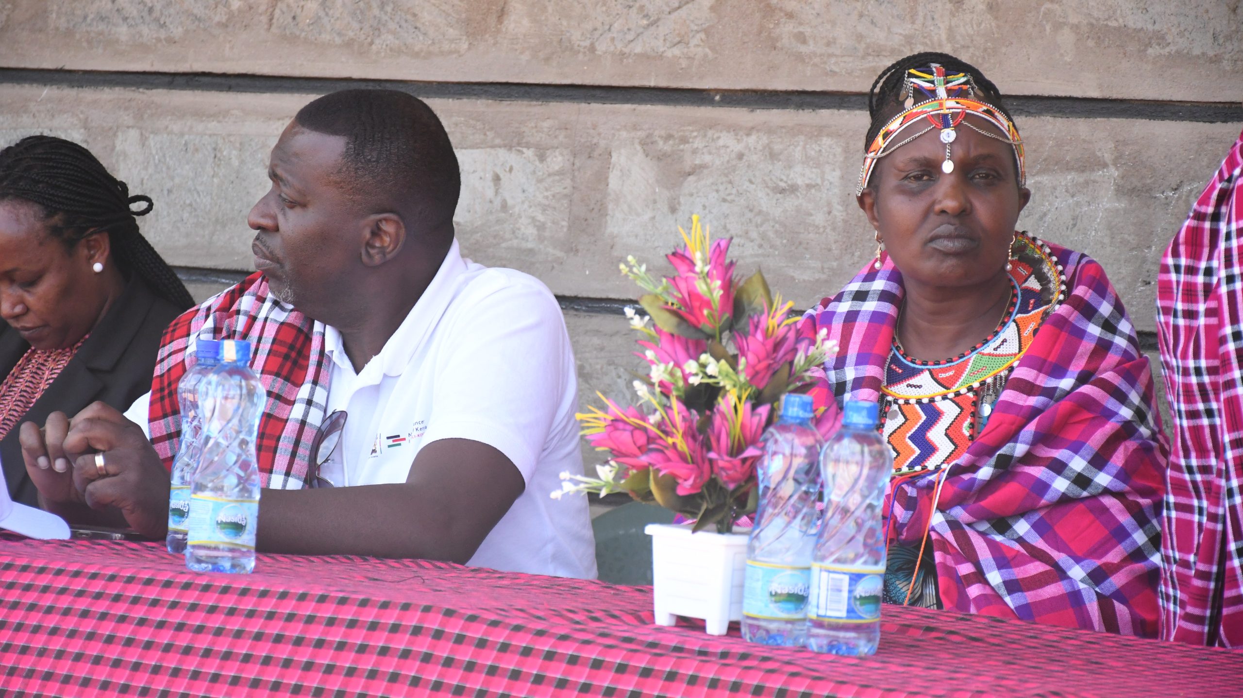
The event garnered significant attention from local leadership, with Hon. H.E Gabriel Lenengwesi, Deputy Governor of Samburu County, and Dr. Lekudere Nassir, CEC for Health, joining the celebration. Hon. Lengerus Pauline, the Women Representative, played an important role in ensuring the event’s success, demonstrating her unwavering commitment to the community’s well-being. Her dedication was crucial in coordinating efforts and ensuring a smooth and impactful launch.
The CS expressed her profound gratitude to the NCI-K team, stating, “I would like to thank NCI-K leadership led by the chair and the CEO for the great work that they have done here in terms of prevention, management, and rehabilitation for patients in the region and also the country.” She further emphasized that the opening of the oncology clinic is a tangible reflection of the government’s unwavering commitment to the treatment and management of cancer.
On her part, Samburu Women Rep, Hon. Pauline Lenguris, echoed the sentiment, expressing her appreciation to NCI-K for making cancer treatment a reality in the county. “This is our first day to start treatment for cancer, and I would like to thank the National Cancer Institute for making this a reality,” she declared.
Speaking during the event, the Cabinet Secretary assured the Samburu County government of the national government’s continued support, particularly in maximizing HPV vaccine uptake. The CS also emphasized the importance of residents registering for the Social Health Authority (SHA) to fully benefit from the oncology package, ensuring that financial barriers do not impede access to crucial cancer care.
The newly launched oncology clinic represents a significant leap forward in addressing the challenges of cancer care in Samburu County. Previously, residents faced long and arduous journeys to access specialized treatment. This clinic will now provide vital services closer to home, reducing the burden on patients and their families.
The launch signifies more than just the opening of a facility; it represents a commitment to providing accessible, high-quality cancer care to all. The collaboration between the national and county governments, along with the expertise of NCI-K, ensures that the people of Samburu County will have access to the resources they need to fight cancer.
This initiative is a beacon of hope, demonstrating the power of partnership in improving healthcare outcomes. As the doors of the oncology clinic open, a new chapter begins, one filled with promise and the potential to save lives.